
Glaucoma
Also known as: Open-angle glaucoma, Angle-closure glaucoma, POAG
Overview
Glaucoma is a group of progressive optic neuropathies characterized by retinal ganglion cell loss and corresponding visual field defects, most often associated with elevated intraocular pressure (IOP). It is a leading cause of irreversible blindness worldwide, and because early disease is asymptomatic, detection depends on structural and functional screening. Lowering IOP remains the only proven, modifiable way to slow progression.
- Type
- Progressive optic neuropathy
- Primary risk factor
- Elevated intraocular pressure
- Only modifiable target
- IOP reduction
- Hallmark
- RNFL thinning, cupping, VF loss
Key clinical points
- Primary open-angle glaucoma (POAG) is the most common form; angle-closure is less common but can be sight-threatening acutely.
- IOP is the principal modifiable risk factor, but normal-tension glaucoma occurs at statistically normal pressures.
- Diagnosis integrates optic nerve appearance, OCT of the retinal nerve fiber layer, and standard automated perimetry.
- Treatment escalates from topical medications (prostaglandin analogs first-line) to laser trabeculoplasty, MIGS, and incisional surgery.
- Because vision loss is irreversible, the goal is early detection and slowing progression, not restoring lost field.
Overview and classification
Glaucoma encompasses several distinct diseases that share a characteristic optic neuropathy with loss of retinal ganglion cells and their axons. It is broadly classified by anterior chamber angle anatomy (open vs. closed) and by cause (primary vs. secondary).
Primary open-angle glaucoma is the most prevalent form in most populations. Primary angle-closure disease, driven by anatomic crowding of the anterior segment, carries a higher risk of acute, symptomatic pressure spikes. Secondary glaucomas include pseudoexfoliation, pigmentary, neovascular, steroid-induced, and uveitic forms.
Signs, symptoms, and risk factors
Open-angle glaucoma is typically asymptomatic until advanced, which is why it is often called a silent disease. Acute angle closure, by contrast, presents with severe pain, halos, a mid-dilated non-reactive pupil, and markedly elevated IOP requiring urgent intervention.
Established risk factors include elevated IOP, older age, family history, African or Hispanic ancestry, thinner central corneas, and higher myopia. Pseudoexfoliation and pigment dispersion increase secondary risk.
Diagnosis
A comprehensive evaluation includes IOP measurement, gonioscopy to assess the angle, dilated optic nerve examination for cupping and rim thinning, and pachymetry for central corneal thickness. Optical coherence tomography quantifies retinal nerve fiber layer and ganglion cell complex loss, often before perimetric defects appear.
Standard automated perimetry documents functional loss and is central to staging and monitoring. Because single measurements vary, longitudinal trend analysis of both structure and function is essential to distinguish true progression from noise.
Management
Lowering IOP is the only intervention proven to slow progression. Prostaglandin analogs are first-line for most patients given once-daily dosing and efficacy; beta-blockers, alpha agonists, carbonic anhydrase inhibitors, and rho-kinase inhibitors are additional classes.
Selective laser trabeculoplasty is an effective first-line or adjunctive option. Minimally invasive glaucoma surgery (MIGS) has expanded the middle ground between drops and traditional filtering surgery, while trabeculectomy and aqueous shunts remain the mainstay for advanced or refractory disease. Angle closure is additionally addressed with laser peripheral iridotomy and increasingly early lens extraction.
Glaucoma videos

Dr. Christine Funke on the 2026 Interventional Glaucoma Masterclass by the Fundingsland Group.

Glaucoma Management
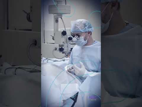
Arkadiy Yadgarov, M.D answers "What excites him the most right now about #glaucoma

How to define severe glaucoma

MTMT and Refractory Glaucoma

Methodical Approach to Teaching #MIGS- Dr. Sahar Bedrood

Hawaiian Eye Recap with Dr. Paul Sign- Glaucoma Drug Delivery

Microinvasive Glaucoma Surgery with Dr. Nicole Bajic
Frequently asked questions
- Can glaucoma be cured?
- No. Glaucomatous vision loss is irreversible, so the goal of treatment is to lower intraocular pressure enough to halt or slow further optic nerve damage and preserve remaining vision.
- What is normal-tension glaucoma?
- Normal-tension glaucoma is glaucomatous optic neuropathy and visual field loss occurring at intraocular pressures within the statistically normal range, underscoring that factors beyond pressure contribute to susceptibility.
- Is selective laser trabeculoplasty a first-line treatment?
- Yes. Evidence supports selective laser trabeculoplasty as a safe and effective first-line option for open-angle glaucoma and ocular hypertension, and it is frequently offered before or alongside topical therapy.
References & further reading
Related conditions
Cataracts
A cataract is any opacification of the crystalline lens that degrades vision, most commonly from age-related changes. It is the leading cause of reversible blindness worldwide and is treated definitively with surgical lens removal and intraocular lens (IOL) implantation. Modern phacoemulsification is among the most frequently performed and successful procedures in medicine.
Clinical overviewRetinaDiabetic Retinopathy
Diabetic retinopathy is a microvascular complication of diabetes mellitus and a leading cause of vision loss among working-age adults. It progresses from non-proliferative changes—microaneurysms, hemorrhages, and exudates—to proliferative disease with neovascularization that risks vitreous hemorrhage and tractional detachment. Glycemic and blood pressure control plus timely screening are the foundation of prevention.
Clinical overview